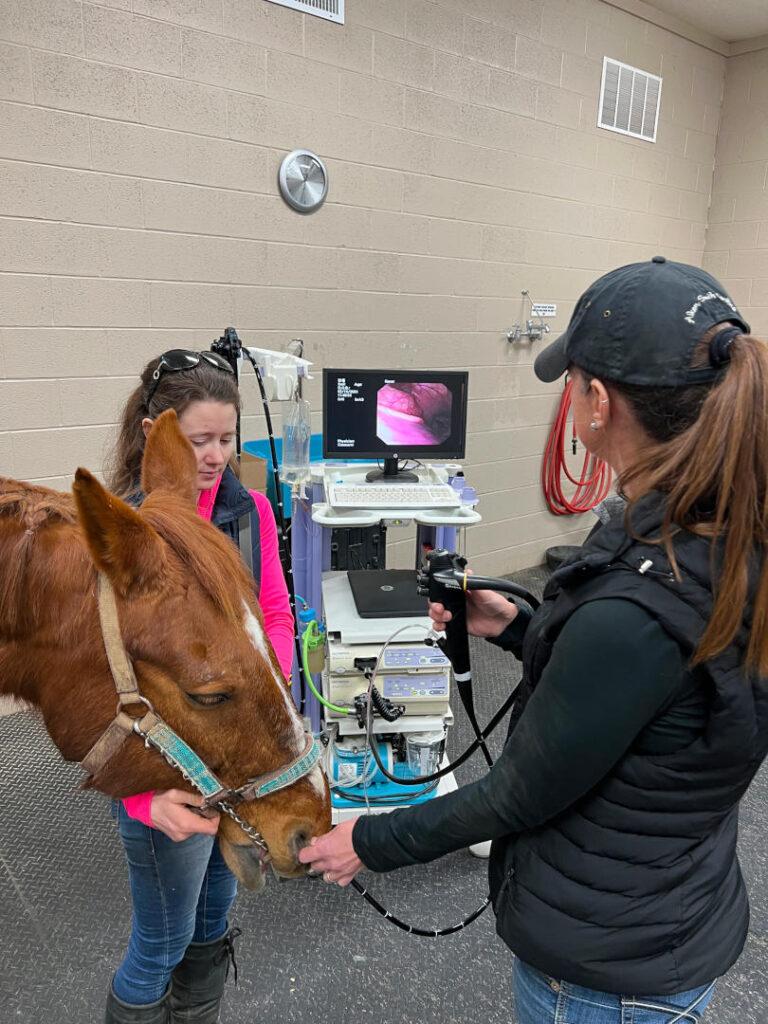
Gastroscopy – During this procedure the veterinarian uses a long flexible video endoscope to help evaluate the esophagus, stomach and even the beginning of small intestine for any abnormalities or disease (ie ulcers, stomach impaction, tumors/growths, etc). This can be done under light sedation and the images can be stored for later analysis and reference for follow up examinations.
Prior to the gastroscope the horse should be fasted for 16-18 hours and water should be removed 2-4 hours prior to the procedure
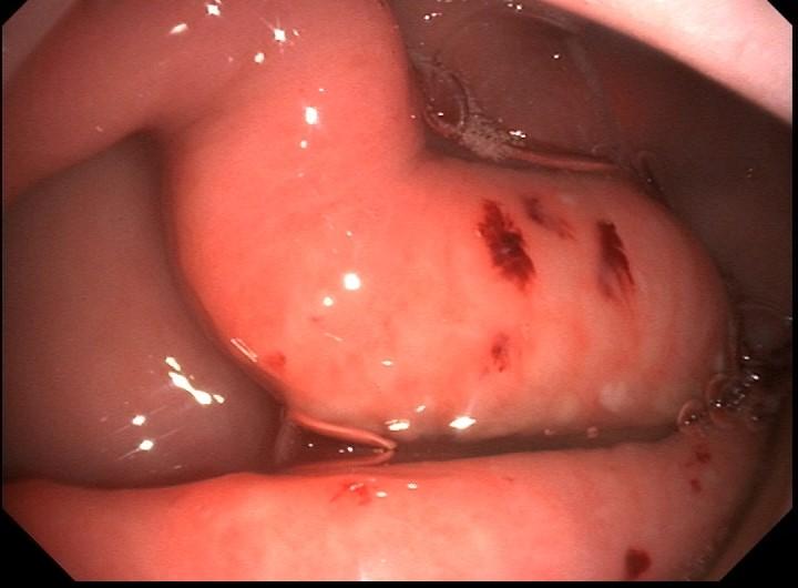
It has been estimated in various studies that anywhere from 50%-100% of performance horses may have some degree of gastric ulcers. Ulcers can appear in various areas of the stomach of the horse. These areas include the squamous (non-glandular area), glandular and margo plicatus. Gastroscopy is the gold standard for diagnosing and fully evaluating the stomach. Depending on the severity of the ulcerations and locations the treatment regiment, time off, and medications can be different. The gastroscope not only helps with the treatment but also allows the veterinarian to track the progress of the horse.
Common signs of gastric ulcers include:
Colic
Weight loss
Change in attitude or behavior
Poor performance
Resistance under saddle
Poor hair coat
Abdominal ultrasound – using digital ultrasound the veterinarian can help evaluate many of the structures of the abdomen including large and small intestine, kidneys, liver, spleen. This procedure can usually be done with little sedation if any. Although there are some limitations due to the size of the horse and interference it is still an important modality.
Abdominocentesis – also known as a “belly tap”, this procedure involves the veterinarian sampling and then analyzing a small amount of fluid taken from the abdomen of the horse. A small area on the horse’s underbelly is prepared and a small amount of fluid is taken using aseptic technique under sedation and local anesthesia. Normally there is a small amount of clearish fluid that helps bath the internal organs. Changes in color, quantity or even presence of feed material can help guide the veterinarian’s diagnosis and treatment plan
Biopsy – if indicated by your veterinarian a biopsy can be taken using a special instrument that can be passed through the video gastroscope itself and sent in for analysis.
Reproduction and Genitourinary
Cystoscopy – using a flexible video scope the horse’s urethra, bladder and urethral openings can be visualized. The procedure is performed with the horse under sedation while standing. Cystoscopy is many times used to evaluate the bladder for tumors and stones or investigate reasons for hematuria (blood in urine).
Urinary catheterization – under standing sedation a sterile urinary catheter can be guided into the urinary bladder of the horse. Some reasons for performing this procedure include obtaining sterile urine sample for analysis, draining the bladder in cases where the horse is not able to adequately urinate or to collect timed volumes to evaluate kidney function.