Categories
Cleveland Equine Clinic News

Potomac Horse Fever

Potomac Horse Fever

By: Chauncey Smith, DVM

Potomac Horse Fever (PHF) is caused by the bacteria, Neorickettsia risticii. Equids acquire the bacteria by consuming infected aquatic insects on pasture. The common culprit is the mayfly. The disease often occurs in late summer and throughout fall, when the insect load is highest. Cases have been reported in the winter and spring seasons. Potomac Horse Fever can affect any horse. 

Owners and trainers might note lethargy and inappetence prior to other clinical signs. Abnormal parameters, such as an elevated heart rate and/or respiratory rate are described. These findings are caused by an elevated body temperature. A fever is any temperature above 101.5F. Potomac Horse Fever will cause diarrhea in most patients. In some cases, horses will develop laminitis. Pregnant mares are at risk of abortion.

Potomac Horse Fever is a medical emergency. Early treatment can reduce or eliminate symptoms. Veterinarians will use nonsteroidal anti-inflammatory therapy to manage the fever. Specific antibiotic protocols have been described for the treatment of this disease.

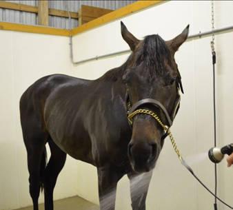
Care providers should note any change in the attitude or appetite of a horse. Having a thermometer and stethoscope is recommended. Any fever should be reported to your veterinarian. A horse with an elevated temperature should be actively cooled via cold water horsing or an alcohol bath. Water intake should be monitored because hydration status can greatly affect the disease process.

A vaccine for Potomac Horse Fever is available. The vaccine does not prevent the disease but can reduce its severity. Horses should be vaccinated prior to the peak seasons of exposure. The clinical signs of Potomac Horse Fever are similar to those of many infectious diseases. Isolation and biosecurity protocols should be implemented if a fever or diarrhea are noted in any horse. Potomac Horse Fever, however, is not contagious so these practices will not be warranted once a diagnosis is confirmed.

Categories
Cleveland Equine Clinic News

Choke in Horses

Choke in Horses

By Chauncey Smith, DVM, Cleveland Equine Clinic

Most chokes are not life threatening. Always withhold feed following sedation. Do not feed a hot horse. Please call a veterinarian if you suspect your horse is choking.

 If you suspect your horse is choking, please remove food and water from the stall. Maintain a quiet environment where the horse can relax. You should contact your veterinarian. Common veterinary treatments for choke include administration of sedatives and relaxants. A nasogastric tube is often passed for manual treatment of the choke. In severe cases, the veterinarian may elect to pass an endoscope to aid in removal of the obstructive material and to assess the esophageal damage. Some horses can chronically choke (choke multiple times). An endoscopic examination may help to determine the cause.

 Severity and longevity of the obstruction can be associated with future complications. Aspiration pneumonia can occur if the material is inhaled in the airway. An esophageal stricture can form due to tissue damage at the site of the obstruction. The stricture can cause clinical signs days to weeks following the initial incident.

 Esophageal obstruction is often seen after feeding. A horse with choke can have saliva and food material present in the mouth and nostrils. That material can instigate a cough if it passes into the trachea. Rigidity of the neck in an extended position is another common finding. Most choke patients are disinterested in food and water.

 Choke is the horsemen’s term for an esophageal obstruction. It is the most common disorder of the equine esophagus. As the horse ages, poor dentition and decreased saliva production can cause incomplete mastication of feed and cause choke. Regardless of age, a horse who has recently been exercised or sedated can be at risk.

Categories
Cleveland Equine Clinic News

Heatstroke in Horses

Heatstroke in Horses

By Chauncey Smith, DVM, Cleveland Equine Clinic

The horse thermo-regulates its body temperature via blood vessels in the skin and sweat glands across the body. Hot and humid weather during summer months can make regulation difficult.

Heatstroke occurs at body temperatures above 105 F. At this temperature, the horse can enter a state of shock and organs can shut down. Heatstroke can cause mental depression; resulting in weakness, collapse, and/or loss of consciousness. When concerned about heatstroke, equine professionals can assess for an increased heart rate and respiratory rate. The horse will have pale and dry mucous membranes with poor refill time. The temperature should be closely monitored. 

Rapid cooling and re-hydration are essential for treatment of heatstroke. Water, alcohol, or cold packs can be used to decrease the body temperature. The horse should be moved to a cool and shaded environment. Oral water with electrolytes should be provided to properly re-hydrate the horse. A veterinarian should be contacted as the horse may require focused care. 

Prevention of thermal insult can be accomplished via early training and preparation. Horses should build stamina during cooler months. Owners should water horses undergoing exercise and travel. Health conditions should be reviewed by veterinarians prior to elevated levels of exercise or high environmental temperatures.

Categories
Cleveland Equine Clinic News

A Veterinary-Farrier Relationship

A Veterinary-Farrier Relationship

WHEN WE THINK OF a horse and rider working together to reach a collective goal, how often do we also think of the grooms, farriers, veterinarians, nutritionists, and others who often play an integral role in the development of that team? The collaboration of each of these professions, and how well they transition from one to the other quite often determines the success of the horse and rider. As a veterinarian, I look out for the welfare and health of the horse, but the frequency with which I see some horses only once or twice per year. When a problem or concern is expressed by the owner or trainer pertaining to the horse’s feet, having a constant dialogue and knowledge of what the farrier has been or is currently dealing with is paramount. In the area where I practice there are many farriers, and although I work with a handful frequently, I am constantly meeting and working with new ones. 

Often the reason for our discourse is due to a problem or a perceived ‘problem’ by the owner/trainer/rider. My goal is to determine what and where the problem is located, and establish as definitive a diagnosis as possible for that situation. I recently saw a middle-aged appendix Quarter Horse gelding that shows English. The owner didn’t think that the horse was moving as well as he had in the past and wanted the horse examined. It should also be noted that the farrier had asked for lateral and DP radiographs of this horse’s A Veterinary-Farrier Relationship BUILDING PARTNERSHIPS hooves prior to the owner expressing concern. The horse appeared to have ‘high-low’ front feet with the RF being the lower hoof, and I was suspicious of a slight convex dorsal hoof angle of both hind feet. 

The horse had its hocks and front coffin joints injected with minimal response, approximately thirty days prior to my examination. On motion assessment, on a hard cement straight line, there was an intermittent RF limb lameness. When lunged on soft dirt footing left, an intermittent LF lameness of Grade1/5 on the AAEP scale was noted, and a Grade 1/5 RF lameness when lunged right. Both front feet were hoof tester negative. Diagnostic analgesia was performed, and significant improvement was noted with a palmar digital nerve block in both front feet. The horse naturally started moving more forward. 

His expression in his head was relaxed, he reached down with his head and neck on the lunge line and started bending around the circle through his thorax and haunches more evenly. Lateral and dorsal-palmar/plantar radiographs were taken of both front and hind feet. The right hoof had a neutral palmar angle of the coffin bone and showed signs of previous or ongoing coffin joint inflammation with bone remodeling to the second phalanx. The left hoof had a two-to-three-degree positive palmar angle of the coffin bone and also had signs of coffin joint inflammation with bone remodeling to the second phalanx. Both hind feet had a one-degree negative plantar angle and no other significant abnormalities noted. The positioning of the coffin bone in all four feet when standing was well trimmed medially and laterally.

The radiographs were sent to the farrier and we discussed the examination and radiographic findings over the phone. The farrier, whom I had never met or worked with previously, shared some great insights. Not only had he been concerned about the hind feet of this horse in the past several trims, but he specifically had wanted to know about the right front as he had noted changes to the hoof capsule over time. His instincts and knowledge of seeing this horse cyclically, and observing that changes were starting to occur, led him to

Having a constant dialogue and knowledge of what the farrier has been dealing with is paramount.

notify the owners. This was pivotal in bringing the horse in for a work-up. Many times, farriers and veterinarians communicate with each other because there is significant pathology, lameness, or injury to specific anatomical structures within the hoof capsule or elsewhere. In this case, proactive communication between farrier and veterinarian enabled the farrier to do his job more effectively and hopefully prevent future pathology or injury from occurring