Computed Tomography (CT) scan is an advanced imaging modality that allows for imaging of a horse’s anatomical structure including bone and soft tissue. Unlike traditional digital radiography, which only allows two-dimensional imaging, CT gives multiple planes of information that can be reconstructed into a 3D image. While digital radiography is an excellent imaging modality, it does have its limitations, especially concerning the head and neck regions. In these regions, it is difficult to interpret radiographic images with certainty, due to summation of bone structure within the image. Radiographs are produced as two-dimensional images while computed tomography is capable of multiple planes of images. With diagnostic imaging produced in this manner, the lesion is more likely to be identified. This allows for faster diagnosis and treatment of disease.
Our Pegaso CT scanner provides higher-resolution images than a conventional CT and uses less radiation. It may also be used to perform fluoroscopy. Fluoroscopy is a continuous X-ray image that can be viewed in real-time. The Pegaso CT scanner can be completed with standing sedation for structures such as the head and neck or under general anesthesia for distal structures.
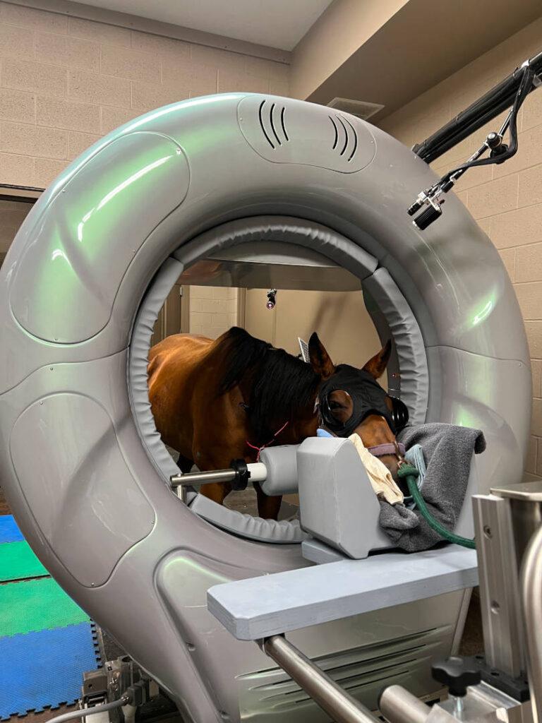
How does it work:
The CT works by rotating around the patient and combining a series of X-ray images taken from different angles. The computer then processes the information to create multiple planes of imaging. These images can then be generated into a 3D image to allow a unique view of anatomical structures. This may allow for a more accurate diagnosis and treatment plan in areas that historically have been difficult to image.
Benefits and uses: