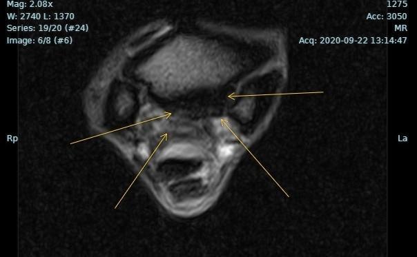
MRI, or magnetic resonance imaging, is a diagnostic modality used to see anatomical structures with more detail than ultrasound and digital radiography alone. MRI is able to visualize bone, soft tissue, and inflammation or fluid accumulation in a detailed and precise location to accurately diagnose and prognose your horse. This in turn allows for improved rehabilitation and treatment options.
The standing MRI (sMRI) uses a 0.27 Tesla magnet and specific technology from Hallmarq to reduce motion and enhance the sensitivity of the imaging. The magnet is able to image from the hoof up to the carpus in front or tarsus behind.
All of these locations are able to be imaged in a standing position, meaning that no general anesthesia is required. Hallmarq rates the image quality of each site’s cases biannually and currently Cleveland Equine Clinic scores a 9/10!
Why refer for an MRI?
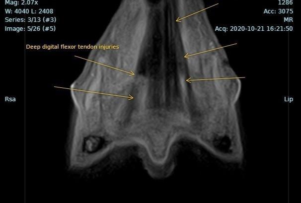
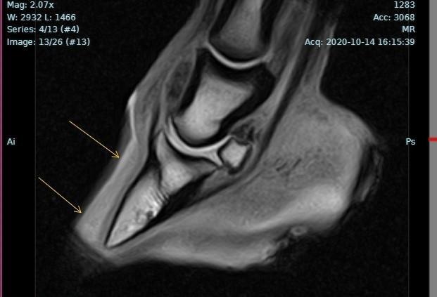
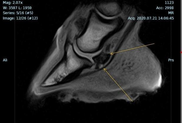
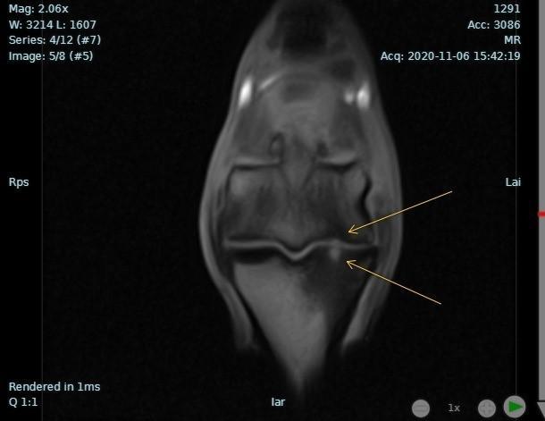
MRI has the ability to visualize lesions or injuries in both soft tissue, bone, and lamina that are not able to be seen on either radiology or ultrasound. Radiographs are good at showing bone, but lag behind in what is really going on in the structure imaged. This is because radiographs show density of minerals within the bones whereas, MRI sees things at a cellular level. Radiographs are not able to show fluid or bone edema/inflammation like a bone bruise, which is a relatively common finding. Ultrasound is very sensitive at visualizing soft tissue and the surface of bone, but cannot visualize within the hoof capsule without extensive hoof preparation and paring of the solar surface, which has its drawbacks. sMRI is able to find small lesions in often hard to visualize areas with ultrasound, like underneath of the ergot, proximal suspensory, the deep digital flexor tendon insertion, and collateral ligaments of the coffin joint.
When a horse presents for various lameness, the most important variable to getting an accurate diagnosis, is having your veterinarian localize the lameness to as specific of a region as possible. For example, your veterinarian initially diagnostically blocks a horse out with a palmar digital nerve block, a block to the mid-pastern region. This is integral to knowing what region of your horse to MRI.
An sMRI will be able to not only provide an accurate diagnosis, but in turn will provide much more accurate information on the duration of time necessary for the horse to heal, if surgery is required, and the prognosis, which may have not been able to be determined previously. Also, and just as importantly, having an accurate diagnosis of a specific anatomical region will allow fluid communication with your farrier to discuss therapeutic shoeing and a long-term shoeing plan for your horse to achieve soundness.
Referral
Cleveland Equine Clinic is a sports medicine and lameness-based practice. Many of the cases that get an sMRI were referred, and we deeply value and appreciate all of the veterinarians that entrust us to take care of their patients. A copy of the sMRI report will be sent to the owner, referring veterinarian and preferably farrier after our doctor directly communicates with you regarding the clinical findings. Should you have any follow-up questions, or require a second or tertiary opinion, please contact our office for assistance.
What happens in the sMRI?
Prior to the sMRI, please have your veterinarian send:
On the day of the sMRI, we do the following: